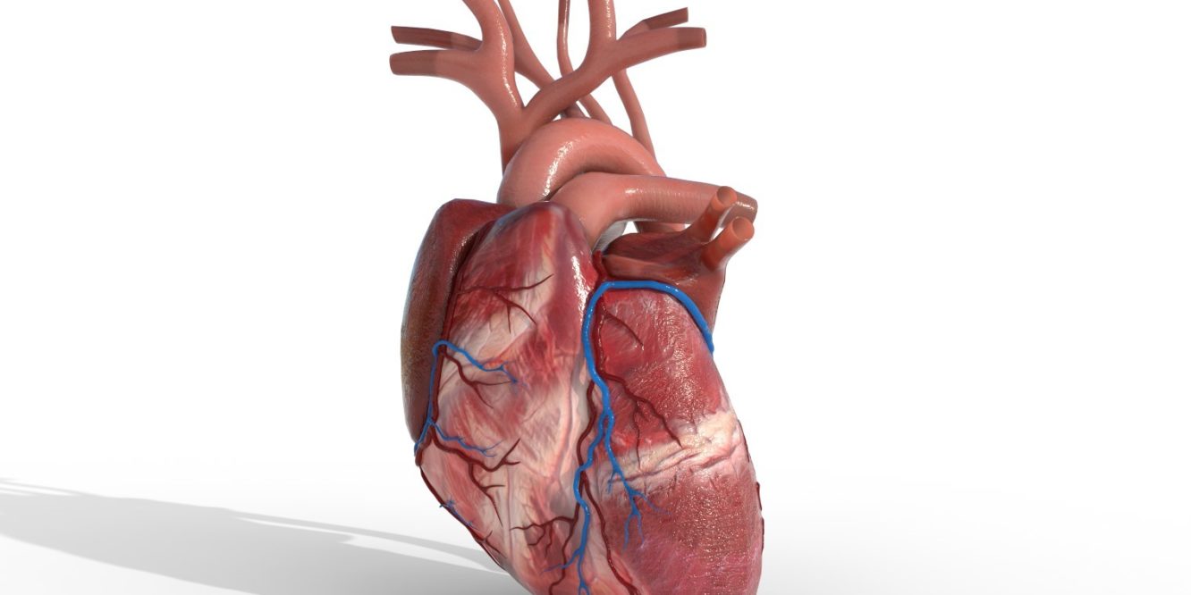
Here we use raw Machine Learning to turn 2D microscopic images of a heart into a dynamical system that allows physicists to easily deal with its related mathematical functions.
This is an example of video semantical extraction which is a modern Artificial Intelligence and Machine Learning tool for extracting semantics from a video. For instance, location, depth, velocity, and detection. We can apply this tech to microscopic images, MRIs, or any 2D, 3D or 4D imaging including live tissue and bio materials.
We have the Machine Learning Algorithms go through a sequence of images to form the fundamental mechanics and dynamics of the system.
We then compute the image displacement between the frames which is a 2D vector field that describes how much displacement and transformation there is between the pixels in one frame to the next. Nothing fancy, just a simple approach using 2D vectors, tensors, and matrices.
We then smooth out the vector field and compute the correlation of displacement vectors where some color or arrow describes the displacement value.
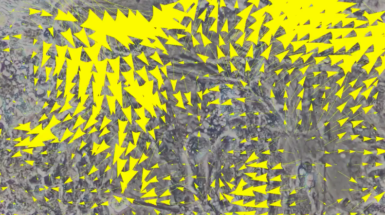
We then do differential geometrical computations on the 2D vector field to compute the divergence. This allows us to compute a scalar map to identify regional tissues on the 2D region to show expansion and contraction.
Red for expansion, Blue for contraction.

We spline the 2D vector field on every coordinate by taking derivatives which allows us to form approximations for differential equations, thus approximating movement in the tissue. Amazingly, by taking the mean norm of the pixels on the screen and building a plot we can model the heart’s pulse as seen on an EKG.
From 2D images we can compute a pulse!